All images on this website have been taken in Leicestershire and Rutland by NatureSpot members. We welcome new contributions - just register and use the Submit Records form to post your photos. Click on any image below to visit the species page. The RED / AMBER / GREEN dots indicate how easy it is to identify the species - see our Identification Difficulty page for more information. A coloured rating followed by an exclamation mark denotes that different ID difficulties apply to either males and females or to the larvae - see the species page for more detail.
Chromista (Fungoids and Yellow-green algae)
'Fungoid' is a term used to describe a species that appears similar to fungi, and was once classed with them, but is now assigned to a different Kingdom - the Chromista.
Some algae-like species are also included in this kingdom in the Phylum Ochrophyta - e.g. the yellow-green algae Vaucheria and Botrydium. [Note that taxonomy is very confusing and subject to change.]
The Phylum Oomycota includes the Albuginaceae (White Blister-rusts) and Peronosporaceae (Downy Mildews) families of species that resemble moulds or mildews. Galls are formed by some species. This group of plant pathogens have a resemblance to fungi, but have cells walls of cellulose or glycan, rather than chitin.
Resources:
- A very good guide to White Blister-rusts and Downy Mildews is available to download from Aber University's website: Chater, A.O., Woods, R.G., Stringer, R.N., Evans, D.A., & Smith, P.A. (2020) Downy Mildews (Peronosporaceae) and White Blister-rusts (Albuginaceae) of Wales.
- See 'Plant Parasites of Europe' website ('Bladmineerders') for images
- Gall descriptions and notes are in Redfern, M. & Shirley, P. (2023) British Plant Galls. FSC
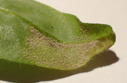
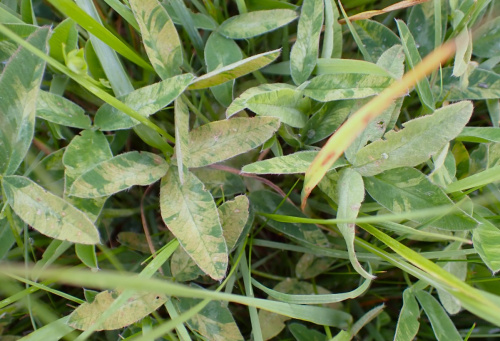
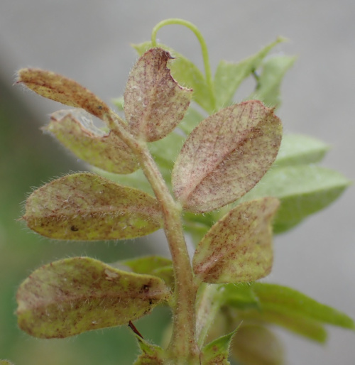
Peronosporaceae - Downy mildews
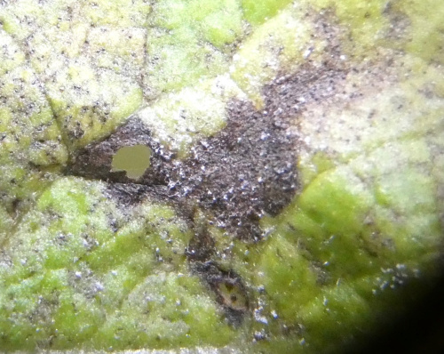
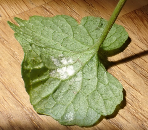
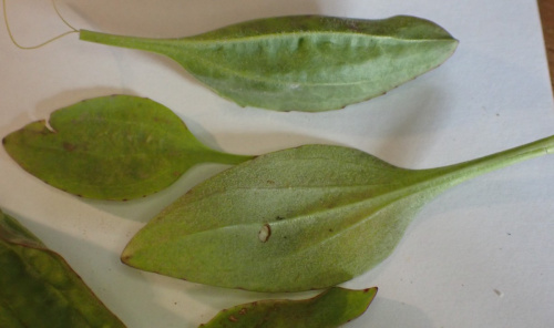
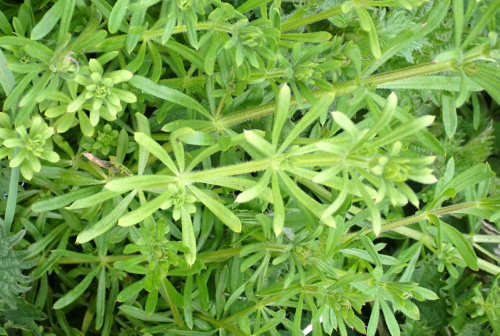
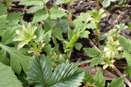
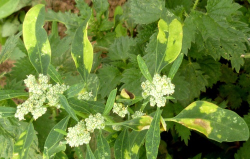
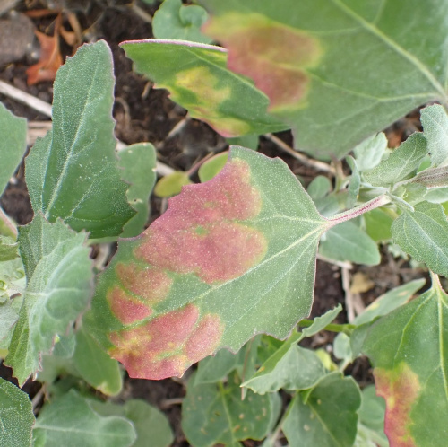
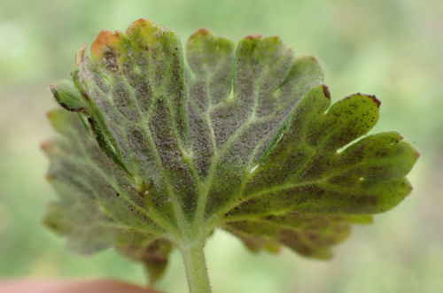
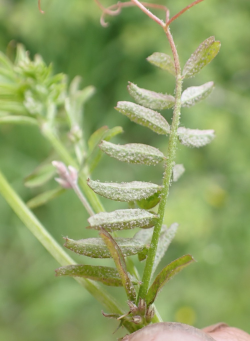
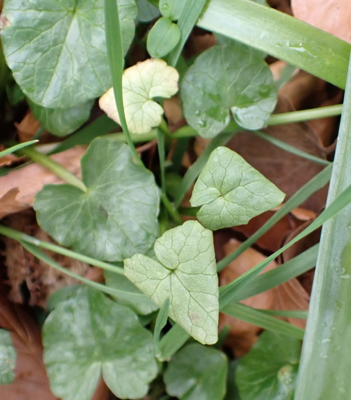
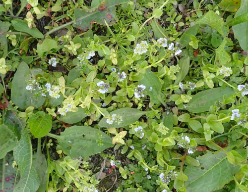
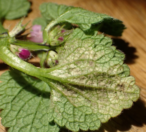
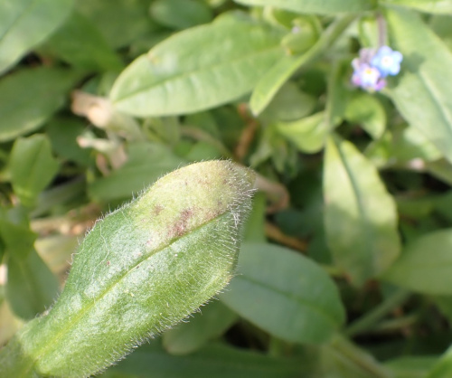
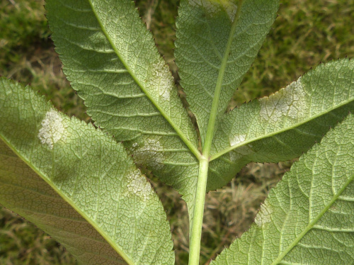
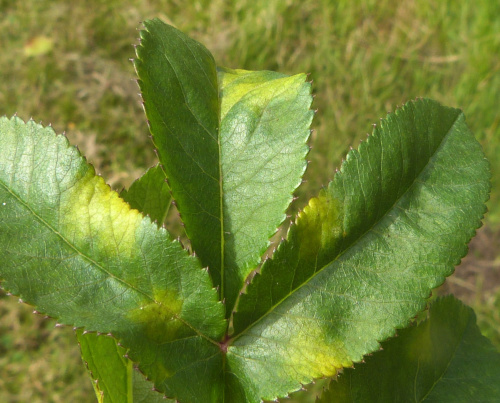
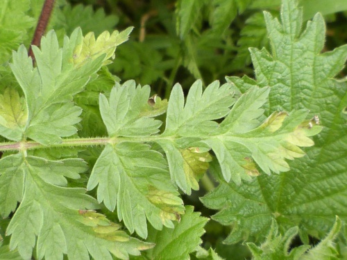
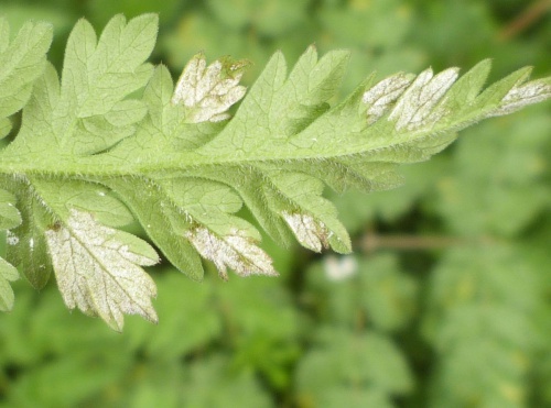
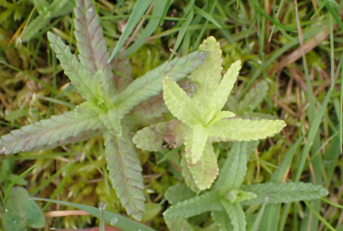
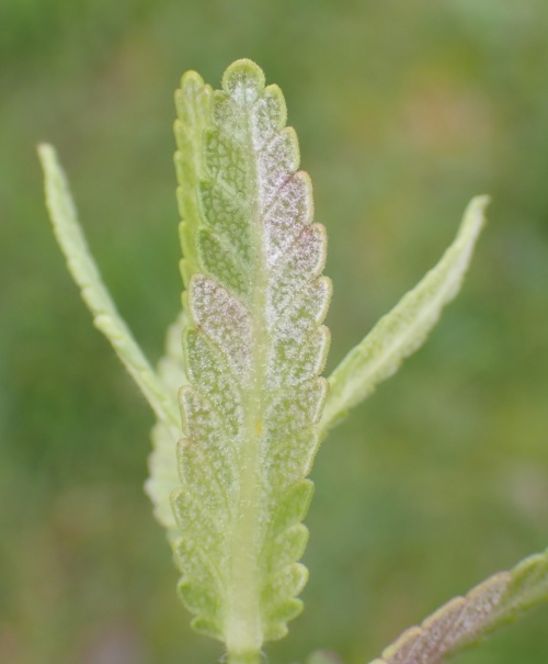
Waste ground, arable margins or damp shady places in hedgerows, woodlands and ditches are often a good place to look. Many species can be seen in the field as a greyish or brownish down or felt on the leaf undersides, often on leaves that are paler, discoloured or slightly thickened, swollen or distorted in some way. Affected plants may be stunted and have leaves with elongated petioles standing erect of the unaffected parts of the plant, or may have yellowish or pinkish blotches on the upper leaf surface. Many are considered to be gall-causers, and are specific to a plant or small range of hosts - it is very important to note the host plant, at least to a genus, when collecting and recording.
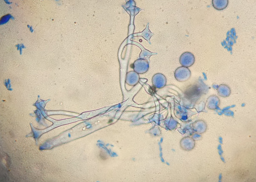
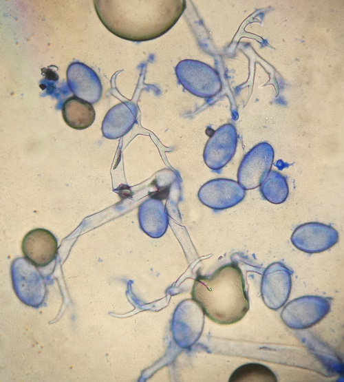
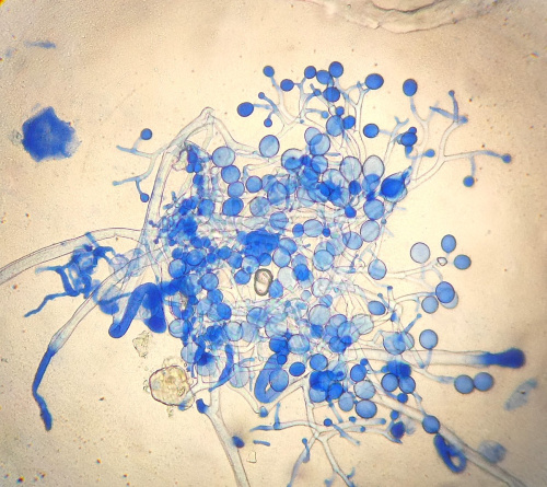
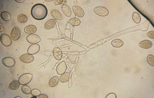
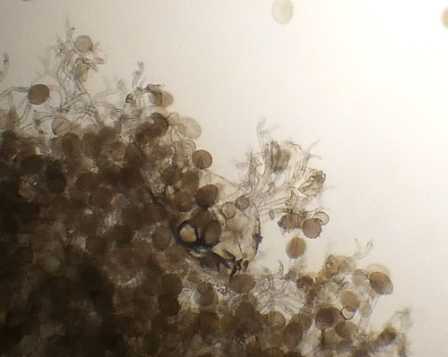
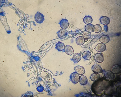
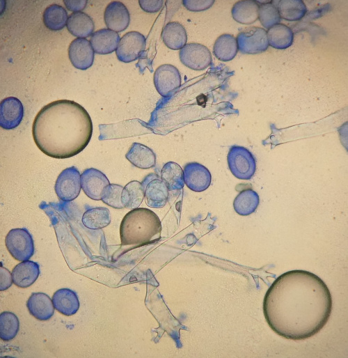
They have a mycelium within the host plant's tissue, which absorbs nutrients from the host. Branching sporangiophores bearing the sporangia emerge from host, usually via the stomata. These tree-like structures are visible under x100 or x400 magnification. A good technique for examining sporangia under the microscope is to press a piece of sticky tape to the mildew, which is usually on the leaf undersurface. The tape is transferred with the mildew to a drop of water on a microscope slide. A small amount of Cotton Blue stain helps to show the branching sporangia.
![]() x400, Cotton Blue stain
x400, Cotton Blue stain
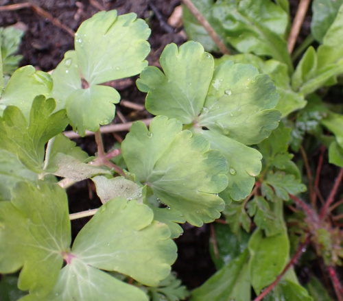
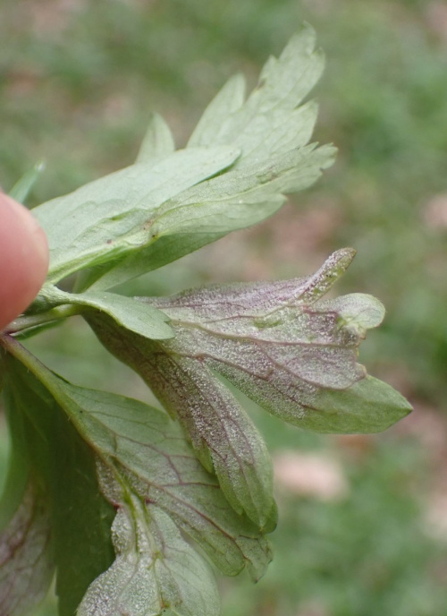
underside of leaves of garden Columbine